The heart is one of the most important and vital organs of the body. It pumps blood, oxygen and nutrients to other organs of the body. Today, due to various reasons including wrong lifestyle and lack of activity, the incidence of cardiovascular diseases is increasing dramatically. The most common cause of death in the world is cardiovascular disease. Worldwide, more people die from cardiovascular disease than from any other cause. Most of these diseases can be prevented and treated by adopting the right lifestyle, reducing tobacco and alcohol consumption, following the right diet, losing weight and increasing the ability to move, the percentage of these diseases can be reduced.
Article Contents:
- Introduction
- CT scan of the heart
1.2. Preparations before a CT heart scan. - Magnetic resonance imaging for imaging of the heart and blood vessels
1.3. Preparations before performing a cardiac MRI. - Echocardiogram
1.4. Types of Echocardiogram.
•Doppler echocardiogram.
•Transthoracic echocardiogram.
•Transesophageal Ejection (TEE).
•Stress echocardiogram.
•Thallium scan (myocardial perfusion scan). - Computed tomography of the heart.
- [Pet Scan] method.
- Nuclear abortion test.
- Nuclear heart scan.
- Final Note
1. Introduction
The most common cardiovascular diseases include stroke or heart attack, heart failure, arrhythmia, heart valve disease, etc. To diagnose and confirm these diseases, we need cardiovascular imaging. Here, a variety of cardiovascular imaging modalities will be explained along with their previous procedures as follows:
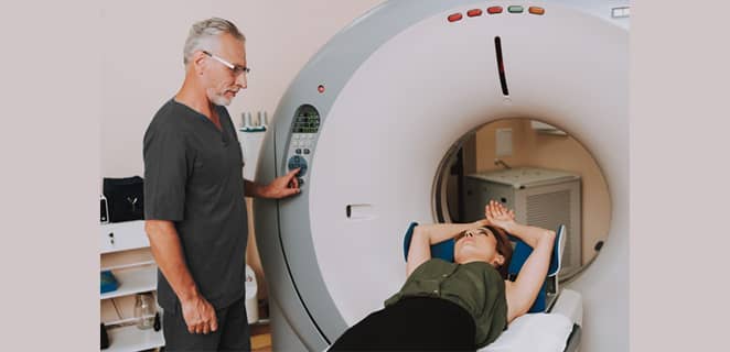
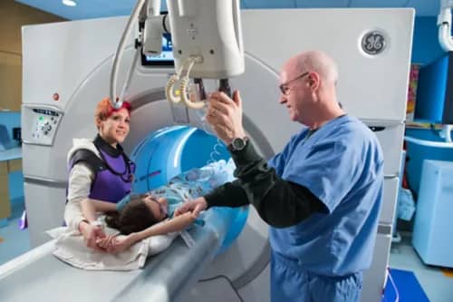
2. CT scan of the heart
Computed tomography is a method of imaging the heart and blood vessels. X-rays are used in this type of imaging. The basis of her work is that she takes pictures of different parts of the body, both horizontally and vertically. His machine uses radiation to take pictures of different parts of the heart and then sends the information to the machine’s memory. Then the computer prepares 3D images of the heart by processing the information. The energy source is a type of X-ray generator.
Computed tomography is used to prepare images of the heart muscle, pericardial sac, coronary arteries, thoracic and abdominal aorta, pulmonary veins, etc. By this, vascular blockages, heart valve problems, congenital heart disease, heart pumping function, heart tumors, etc. are determined.
2.1. Preparations before a CT heart scan
You must bring a prescription for the medicines you are taking. In addition to prescriptions, bring your medical records with you. All kinds of metal items, watches, necklaces, etc. should be left. Most CT scans are done with an injection of contrast material, so be careful not to fast. In a type 64 CT scan, the patient must be given medication to lower the heart rate. To continue taking the medication or to stop taking it before the CT scan, consult your doctor. You should not smoke for at least two hours and avoid tea and coffee for 12 hours. Also, do not exercise before 4 hours and avoid excessive movements. A comfortable dress with an open front is also essential.
3. Magnetic resonance imaging for imaging of the heart and blood vessels
3.1. Preparations before performing a cardiac MRI
If imaging requires injections, fasting should be observed. Bring your medications with you. You must not bring any kind of jewelry, metals or electrical appliances. If you are pregnant, be sure to tell your doctor. Use headphones to avoid being disturbed by the high volume. When you are inside the machine, you must not move. Tell your doctor if you have anxiety or agoraphobia.
Cardiac MRI is the same as MRI and is one of the most important and widely used diagnostic methods. It is used to diagnose diseases throughout the body. This method has many advantages over the other methods we have mentioned. This method is very sensitive to diseases and disorders that are not detected by other methods well defined here. An MRI does not use ionizing radiation and is less harmful than a CT scan. It is a non-invasive method and blood flow imaging is also done.
Biohazards have not been recorded in this imaging method. Also, for soft tissue detection, it has a high accuracy compared to other methods. This procedure is not performed for patients who are afraid of dark or enclosed spaces. Its time is longer than other methods and it cannot be performed in some diseases with batteries and heart transplantation inside the body.
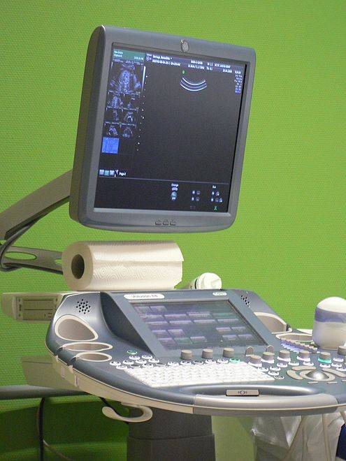
4. Echocardiogram for imaging of the heart
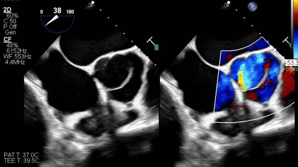
An echocardiogram is a method of imaging the heart and blood vessels in which images of the heart are prepared using sound waves. In fact, it is a kind of high-frequency ultrasound. The resulting images are much clearer than X-rays. It is used for recognizing heart diseases, monitoring heart cavities, their thickness and size, monitoring heart valves and their tightness, diagnosing pericardial infection, diagnosing atrial fibrillation, diagnosing heart murmurs and hypertrophic cardiomyopathy, etc. It is a non-surgical method.
4.1. Types of imaging echocardiogram:
- Doppler echocardiogram: This is used to check blood flow to the heart’s vessels and valves.
- Transthoracic echocardiogram: This is one of the most common types. It is also used for the health of the fetus.
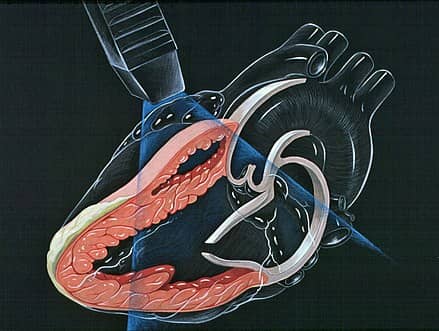
- Through the esophagus (TEE): In this method, the transducer goes down the esophagus and takes an image from where it is closest to the heart.
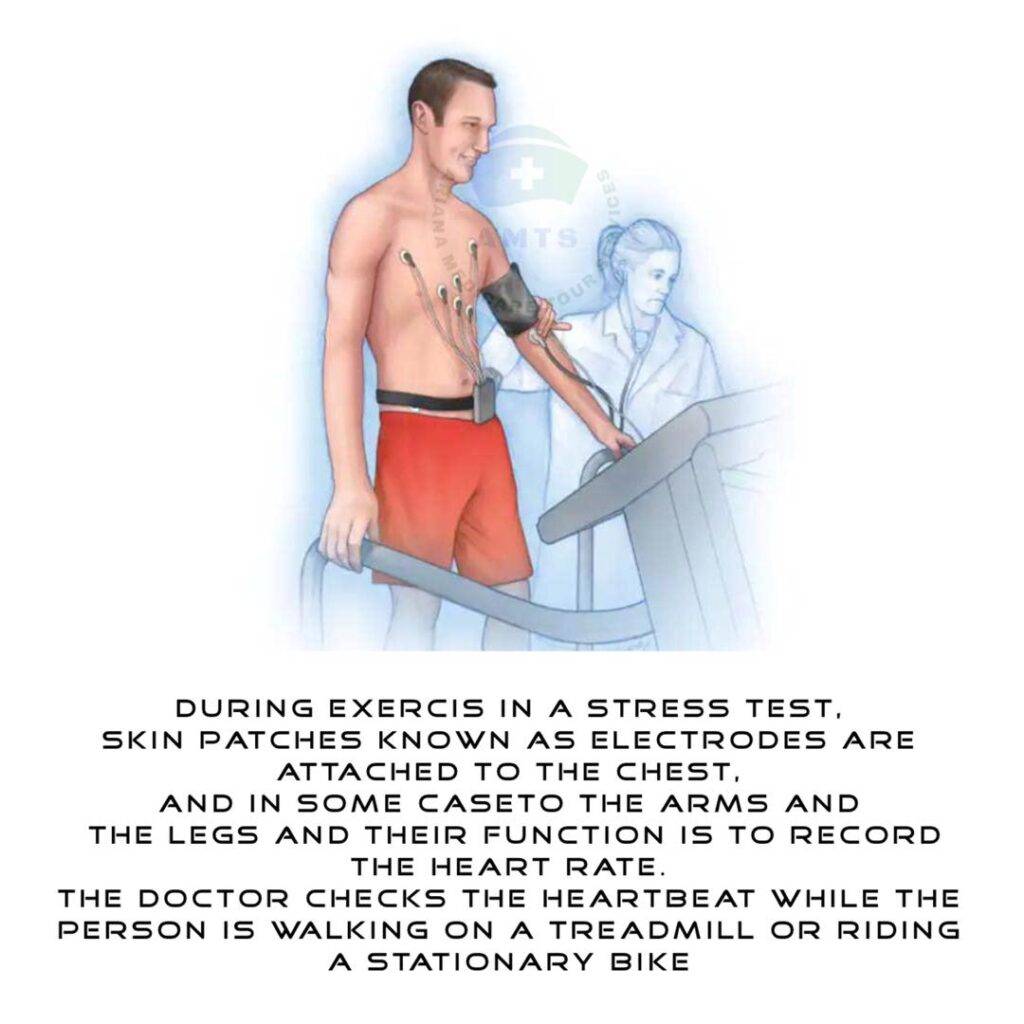
- Stress echocardiogram: In this method, pictures of the heart are taken before and after exercise to check for decreased blood circulation.
- Thallium scan (myocardial perfusion scan)
In this type of imaging, thallium is injected into a patient’s vein and a camera moves around the patient’s body. For this reason, the state of the myocardial blood supply is checked.

5. Computed tomography of the heart
Cardiac CT angiography is also a type of X-ray that is used to check and find blocked pathways. In this test, a contrast material is injected into the diseased arteries, and in this way the blocked pathways are found.
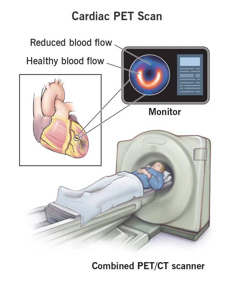
6. PETSCAN METHOD
This imaging method is a combination of CT scan and positron emission tomography. With it, they learn which parts of the heart are not supplied with blood.
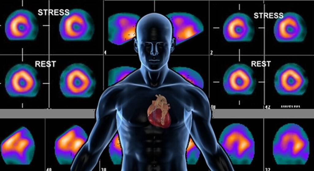
7. Nuclear stress test
By injecting a radioactive substance into the blood and imaging with gamma rays, the function of the heart is checked both at rest and in the active state.
8. Nuclear heart scan
Please make the necessary preparations before having any kind of cardiovascular scan and inform your doctor if you are taking any kind of medication.
9. Final note:
In order to be able to contact the cardiologist more easily, you can use the online consultation, email and WhatsApp.
Sources:
Read more about other articles in the medical fields (Cosmetics and Medical)
- Hair Transplant in Iran
- Rhinoplasty (Nose Job in Iran)
- Face Lift Surgery (Rhytidectomy) in Iran
- Breast Lift in Iran
- Chin Surgery in Iran
- Liposuction in Iran
- Tummy Tuck (abdominoplasty) in Iran
- Butt augmentation (Brazilian butt lift) in Iran
- Breast Augmentation in Iran
- Eye Care in Iran
- Cataract Surgery in Iran
- Diabetic Retinopathy Treatment in Iran
- Heart Surgery in Iran
- Knee replacement in Iran
- Eyelid Surgery In Iran
- Safe Abortion in Iran